Retinal Tears: description and symptoms
Retinal tears usually occur when the vitreous pulls away from the retina and an abnormally strong vitreoretinal adhesion pulls a break in the retina. Tears typically have a U-shaped or horseshoe configuration. The most common symptom is multiple small floaters in the patient’s field of vision, and flashing lights (photopsia). The most concerning symptom is an enlarging, peripheral visual field defect, which suggests that the retinal tear has progressed to a retinal detachment.
Retinal Tears: treatment
Acute tears usually require treatment (retinopexy) with laser or cryotherapy to reduce the risk of retinal detachment. The retinopexy or cryotherapy aim to create a focal scar that sticks down the retina to the underlying retinal pigment epithelium. This prevents egress of vitreous fluid though the retinal break and under the retina, which otherwise could start the process of retinal detachment.
Longstanding tears sometimes lead to a healing response that creates an adhesion between the retina and the underlying RPE (retinal pigment epithelium), creating much the same effect as retinopexy – in this scenario retinopexy may not be required.
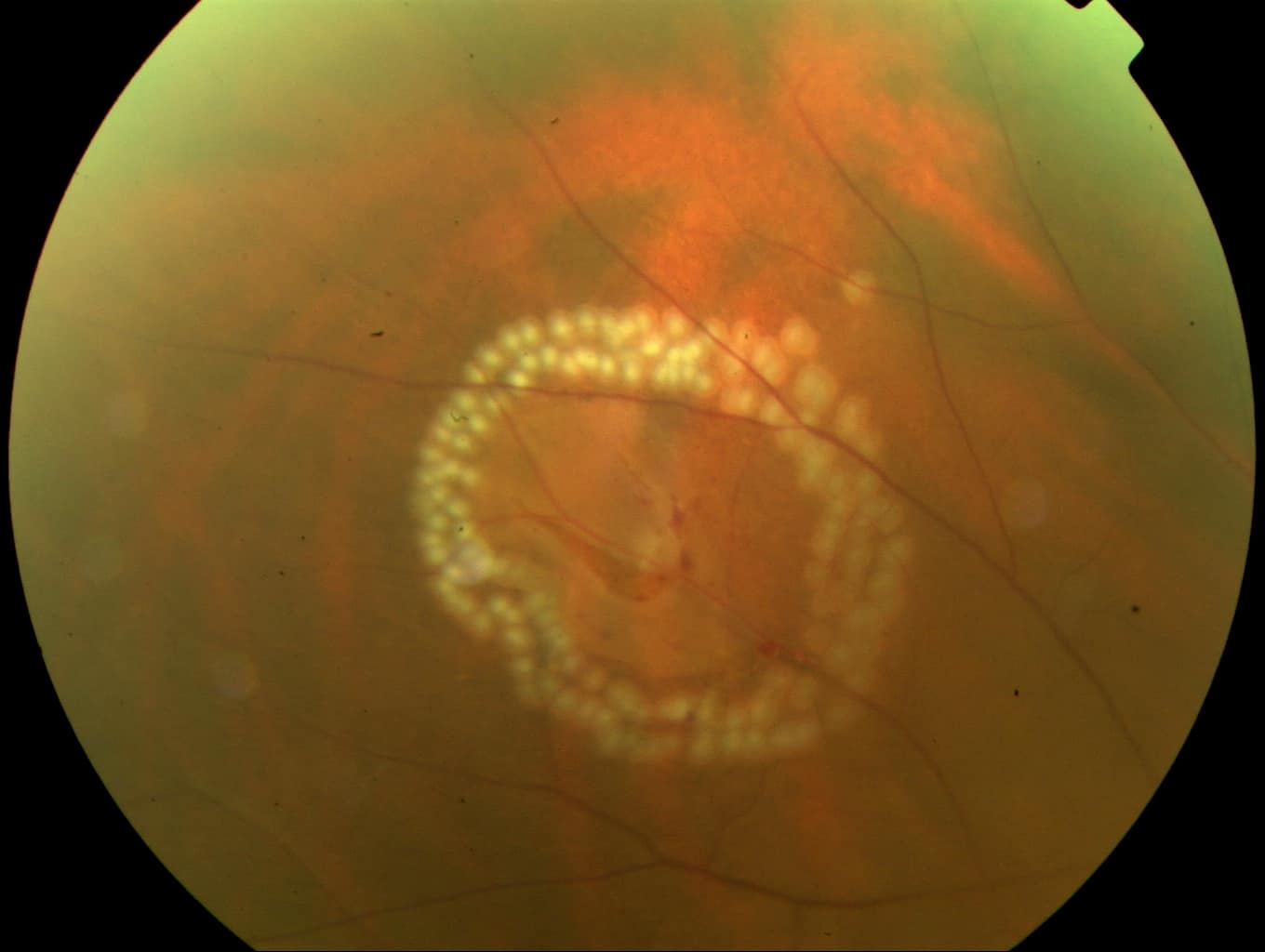
A retinal tear is seen surrounded by recently applied laser retinopexy spots. With time each laser spot will evolve to form a focal scar, promoting adhesion of the retina sealing the break and reducing the risk of retinal detachment.
Retinal holes
Retinal holes have a different pathogenesis – they are round atrophic defects in the retina that can occur without posterior vitreous detachment. They may occur in eyes with lattice degeneration, a thinning of the retina often associated with myopia. In the past they were usually treated, but this is becoming less common.
Referral guide
Symptomatic retinal tears should be seen by an ophthalmologist the same day, or otherwise the next morning. Please call urgently.
Please refer asymptomatic retinal tears picked up during routine optician examination semi-urgently (1-2 weeks).
Atrophic round holes picked up during a routine optician examination can usually be referred routinely.
Provide all patients with a retinal detachment warning, advising them to present immediately if they develop photopsia, floaters, or a visual field defect.
Patient information
Further information about retinal holes and tears and the risks and benefits of treatment can be found in the patient information leaflet.
