Retinal vein occlusion is a thrombosis of the veins that drain the inner retina. It can be divided into branch retinal vein occlusion (BRVO) or central retinal vein occlusion (CRVO) depending on which vessels are involved. CRVO produces haemorrhage throughout the retina, whereas BRVO produces focal involvement.
Symptoms
Symptoms vary according to the area of retina involved, and most importantly, whether or not the fovea is involved. If the fovea becomes ischaemic or oedematous, then visual acuity is affected.
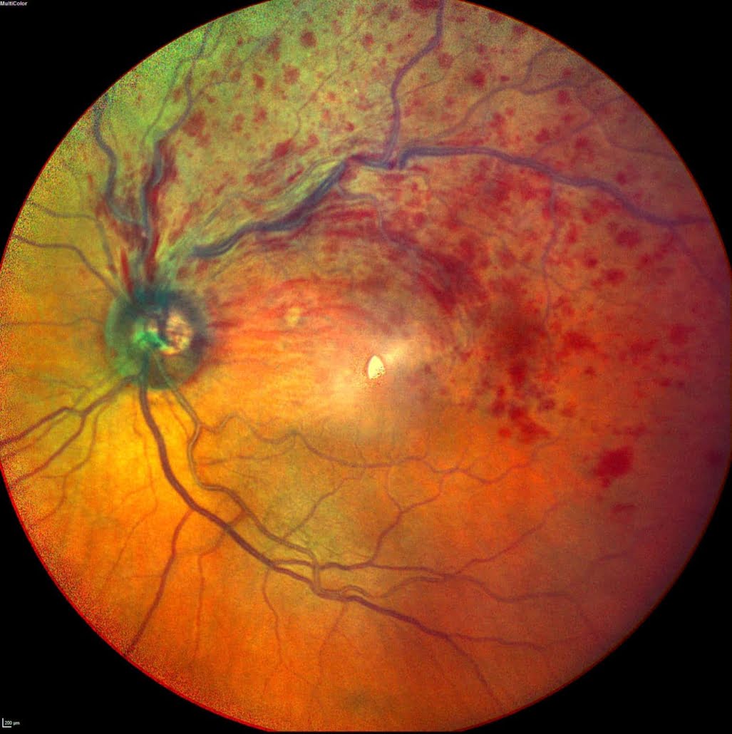
The colour fundus photograph shows haemorrhage due to retinal vein occlusion affecting the top half of the retina.
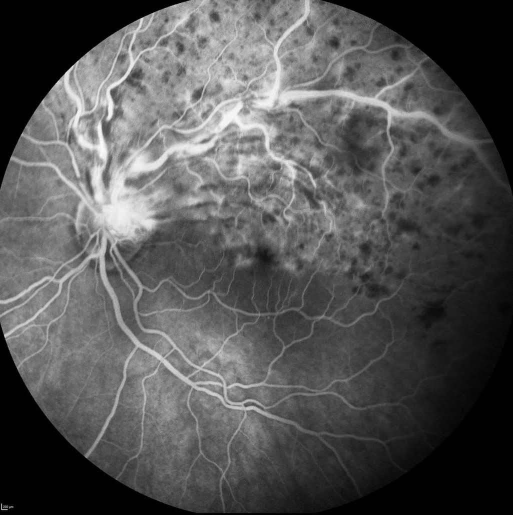
Corresponding fluorescein angiogram showing hyperfluorescence of damaged and leaky retinal blood vessels
.
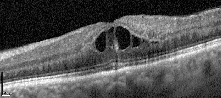
An OCT showing dark central fluid cysts (macular oedema) causing thickening of the central macula.
Normal OCT.
Management
BRVO is initially managed by observation alone, but if macular oedema develops the most common treatment is intravitreal injections of an anti-VEGF drug (Avastin, Eylea or Lucentis). Alternatively, macular grid laser or intraocular steroid implants may be appropriate.
BRVO and CRVO may lead to the development of new vessels that are often treated by panretinal photocoagulation (PRP laser). Neovascularisation is usually a response to extensive retinal ischaemia.
CRVO macular oedema does not usually respond to grid laser, so anti-VEGF drugs are often required, or less commonly, intraocular steroid implants.
Patients diagnosed with vein occlusion should have their cardiovascular risk factors carefully managed to reduce their chance of recurrence or other vascular events. Professor Jackson will usually defer to the patient’s GP for help with this. It is usually advisable to check Full Blood Count, Lipids, ESR or CRP, rule out diabetes and check blood pressure.
Referral guidelines
Please aim to refer patients with acute retinal vein occlusion fairly quickly, with a view to getting an appointment within 1-2 weeks. Please review their cardiovascular risk factors in parallel with the referral.
More detailed information about Retinal Vein Occlusions can be found in the following patient information leaflet.


