What is a retinal vein occlusion?
The retina is the light-sensing layer of cells inside the back of the eye, like the film in the back of a camera. It is supplied by fine arteries and veins. Sometimes the veins become blocked. This blockage can affect either a single branch of veins supplying just one part of the retina (branch retinal vein occlusion) or the central vein supplying the whole retina (central retinal vein occlusion).
The blockage of veins can cause:
- ischaemia – loss of blood supply to the retina
- macula oedema – fluid leakage from damaged veins
- neovascularisation – the growth of abnormal new vessels inside the eye
What are the symptoms of retinal vein occlusion?
Sometimes a small branch occlusion can cause no symptoms, but if the most visually sensitive part of the retina is affected (the macula), or if there is a central retinal vein occlusion, vision is reduced. This usually comes on suddenly and may worsen over a few hours or days. Any vision loss may be mild to severe.
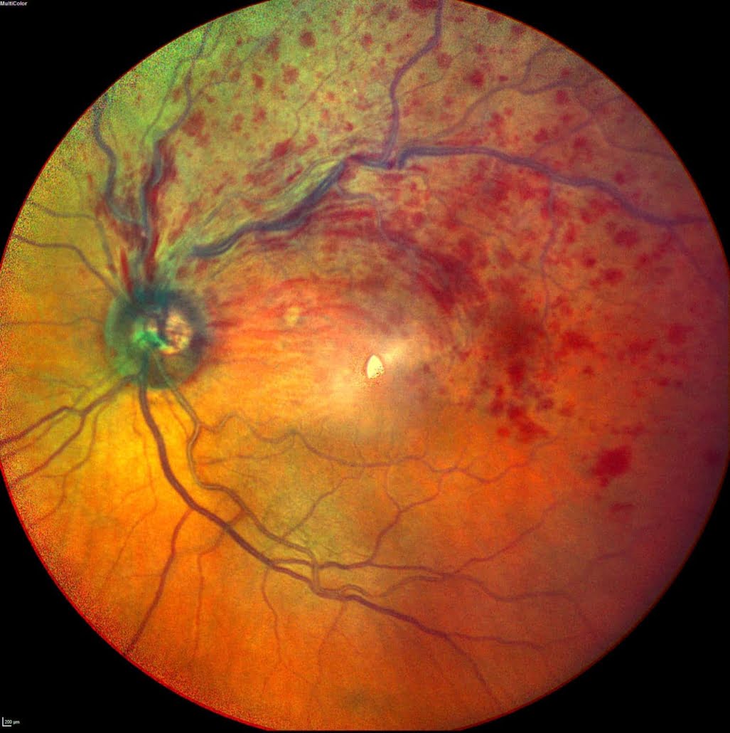
The colour photograph shows the inside of the back of the eye, with haemorrhage due to retinal vein occlusion affecting the top half of the retina.
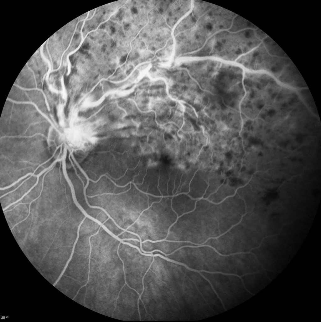
The black and white photo shows damage to the blood vessels in this area using fluorescein angiography.
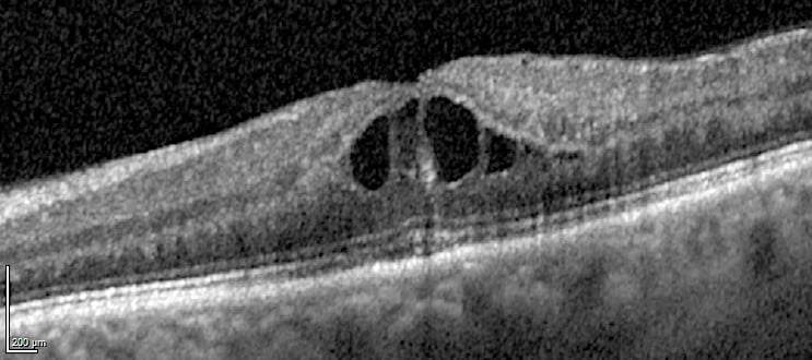
An OCT showing dark central fluid cysts (macular oedema) causing thickening of the central macula.
Normal OCT.
What are the treatments for retinal vein occlusions?
Unfortunately there are no treatments for ischaemia.
The following treatments are available for macular oedema and neovascularisation:
- Injections of an anti-VEGF drug – these may need to be repeated
- Injections of a steroid drug – these are longer lasting than anti-VEGFs
- Laser treatment – either macular laser or pan-retinal photocoagulation
Professor Jackson will discuss the best treatment option(s) for you.
More detailed information about Retinal Vein Occlusions can be found in the patient information leaflet.


